Saastamoinen Foundation – a global partner to art and science. We support artists and researchers on a long-term basis and promote their networks around the world. We facilitate innovative encounters and hold a dynamic art collection. Together we strive towards a better future for next generations.
NEWS
FOUNDATION
Saastamoinen Foundation is a significant supporter of art, science, and societal projects in Finland and internationally. Our values – transparency, internationalisation, expertise and continuity – shape the support we offer to artists, ground-breaking scientific projects, art institutions and community projects aimed at young people.
Partnerships
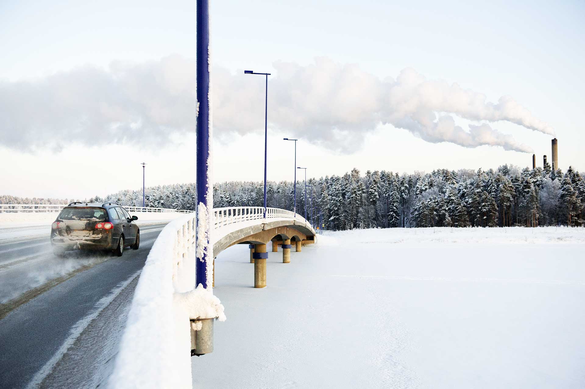
Saastamoinen Foundation operates across the areas of economic, cultural, environmental, and social responsibility.
The Foundation seeks to change the world on the level of beliefs, values, ideals, and worldview. It also promotes transformation in systems and structures, behavior, and technology.
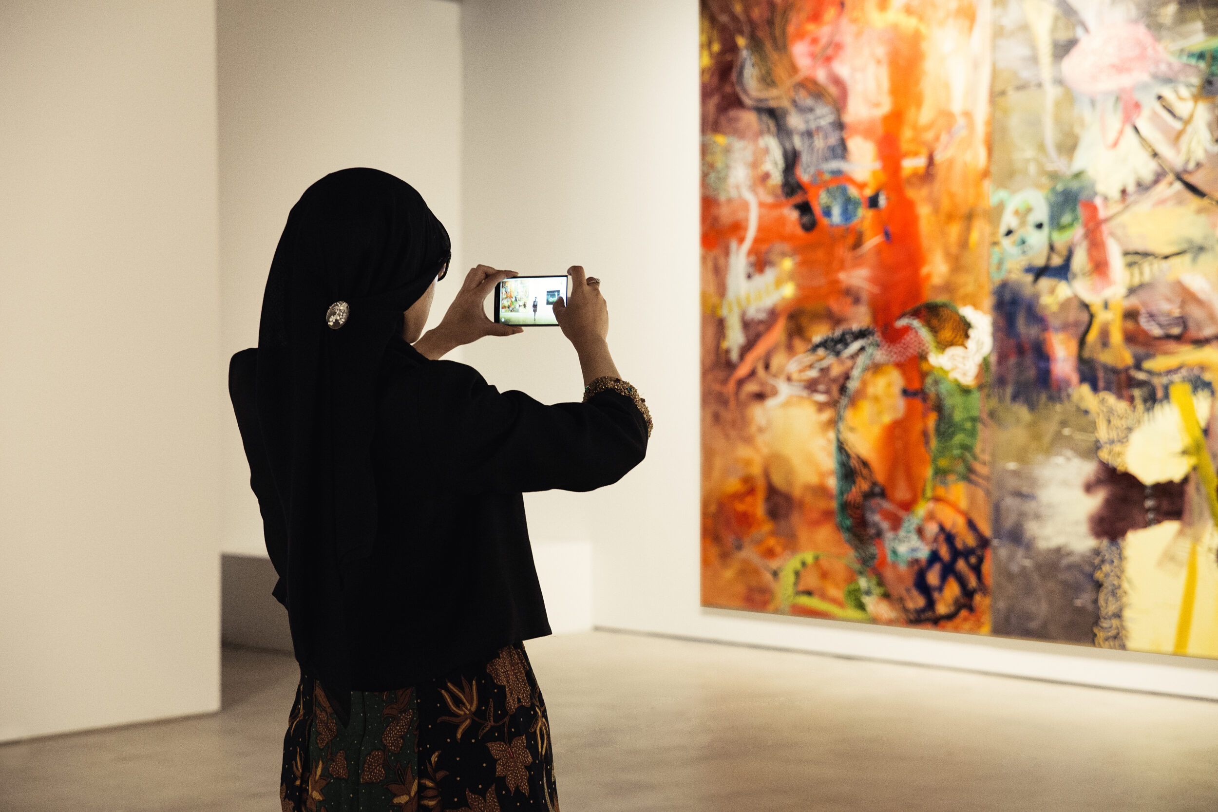
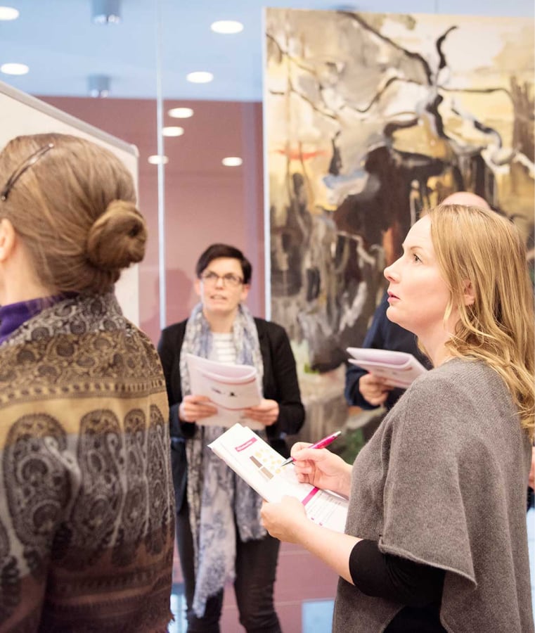
Art
We support art projects of various scales, with a focus on visual art. The art funded by the Foundation fosters curiosity and creativity. Creativity enables us to imagine better futures, develop new solutions, and build meaningful connections.
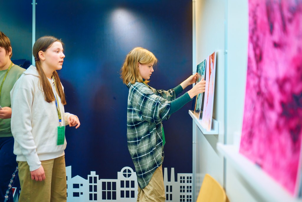
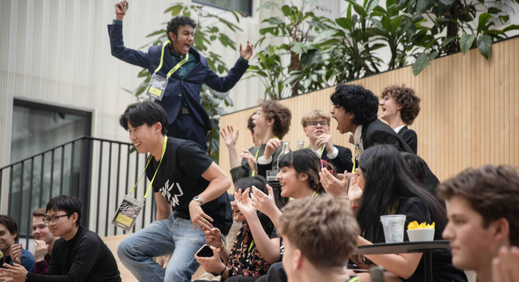
Youth and society
The Foundation supports social initiatives aimed at children and young people. Through our support, we ensure that young people's potential is not wasted—and that they take pride in what they can do.
Art Collection
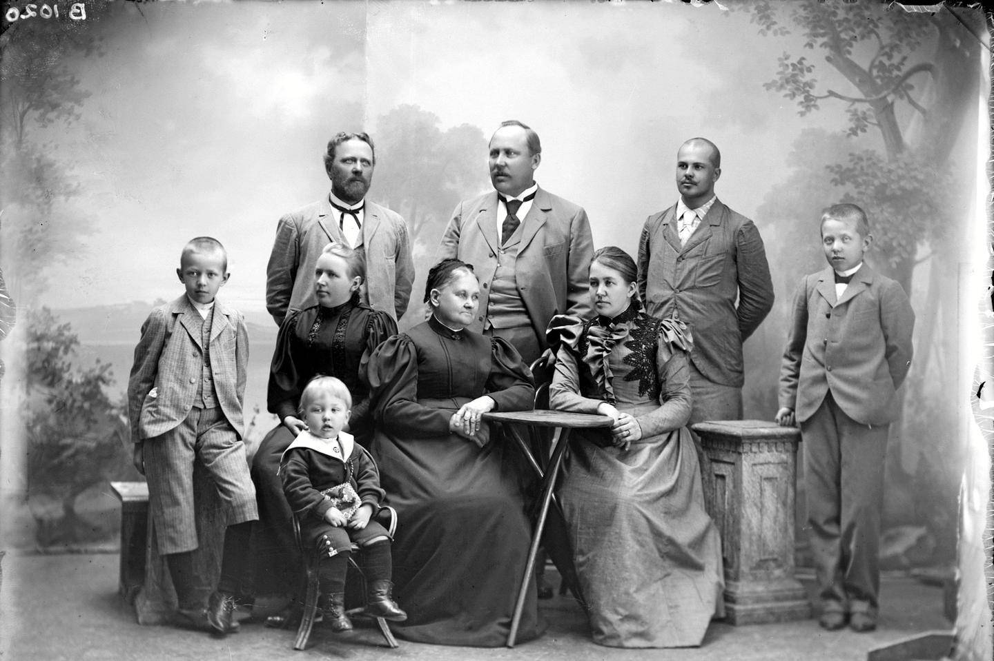
Saastamoinen Foundation is a founding member and long-standing partner of EMMA – Espoo Museum of Modern Art. The Saastamoinen Foundation Art Collection is housed at EMMA, where the works are permanently on display with regularly updated presentations. The Saastamoinen Foundation Art Collection is one of Finland’s most significant collections of Finnish and international art, comprising nearly 3,000 works. The growing and evolving collection builds a bridge between the past and the future.
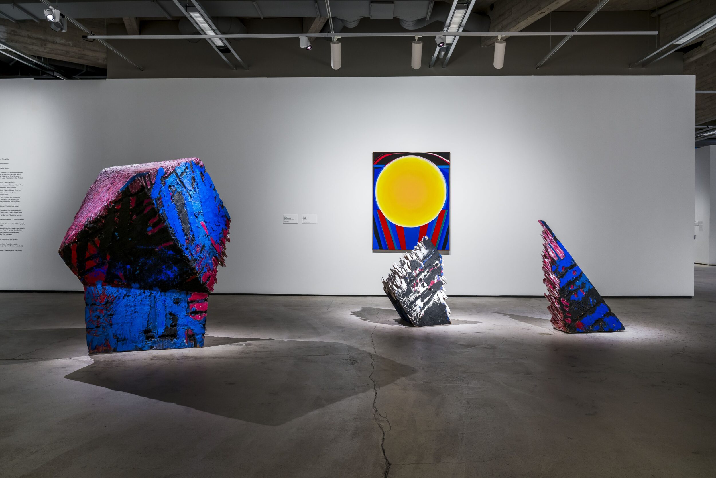
Collection exhibition Dialogues
The collection exhibition Dialogues presents current Finnish and international contemporary art from Saastamoinen Foundation.

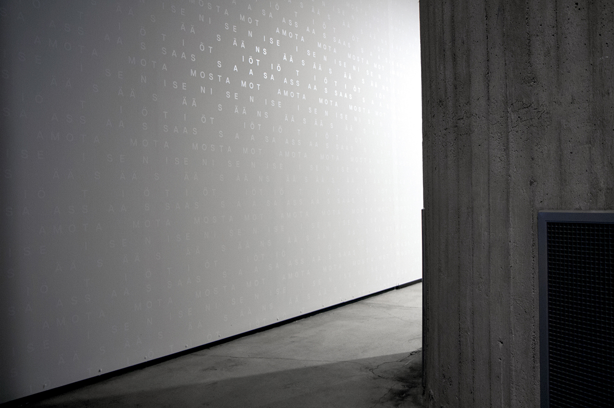
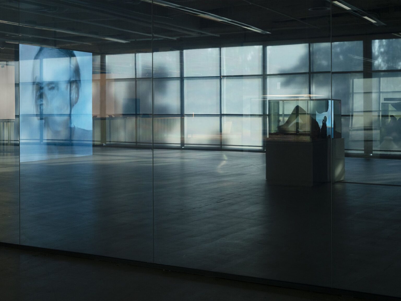
Moving image
Moving image is one of the focus areas of the Saastamoinen Foundation Art Collection strategy. Works in this medium are presented at EMMA as part of the Foundation’s collection exhibition, as well as in a dedicated media art space.
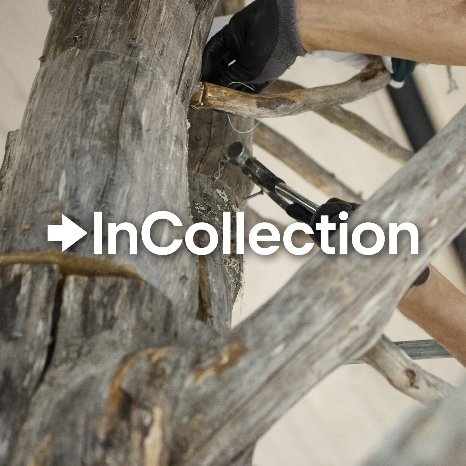
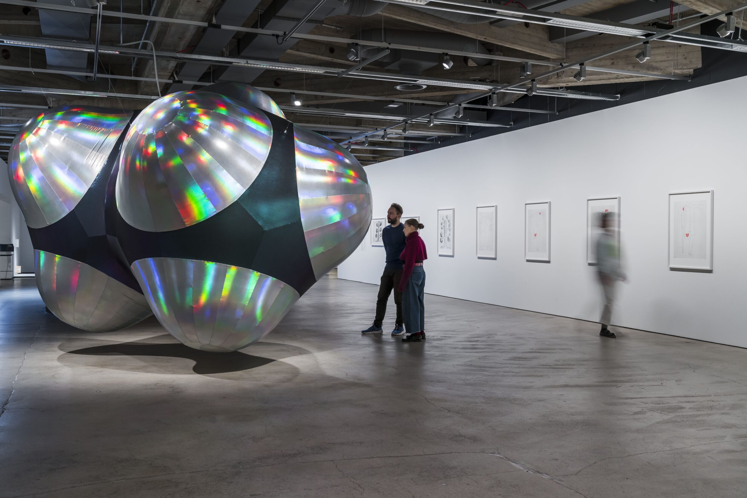
Photo: Antti Laitinen. © Ari Karttunen/EMMA.
Antti Laitinen:
Chiming Forest
Chiming Forest
Antti Laitinen (b. 1975) is an artist based in Somero, Finland, who makes art from materials drawn from his immediate surroundings. In recent years, he has focused on transforming, dismantling and reassembling trees and forest landscapes in his work.
Chiming Forest (2025) is a kinetic installation – an artwork involving movement – that invites us to consider humanity’s capacity and means to shape the natural world. The work is constructed from wood sourced throughout southern Finland, including brushwood left behind in logged areas and root systems of trees felled by wind. These fragments of trees are reassembled into new, hybrid forms and arranged into a forest-like composition – an artificial landscape animated by mechanically produced motion and sound.